In order to better evaluate drug treatments or understand biological processes working directly with whole-slide tissue images, life-scientists need to have immediate and easy access to their image collection independently of the slide-scanning work station.
Furthermore tailoring the workflow and carrying out collaborative and multidisciplinary studies with powerful annotation and quantification tools can help scientists to achieve their research goals faster.
A fully documented open-source solution
Cytomine is driven by its own scientific community
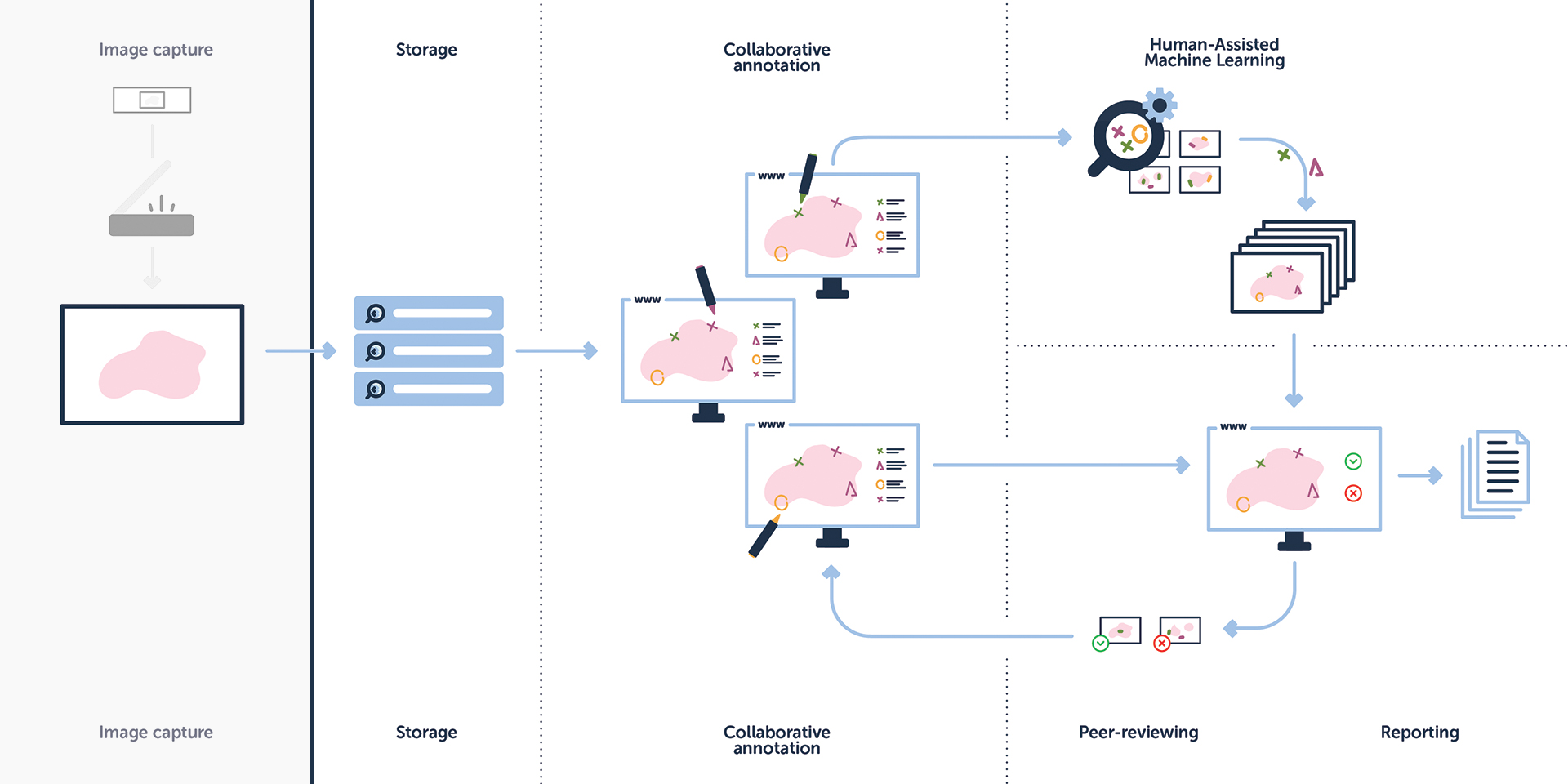
Storage
upload and archive your whole-slide imaging data in Cytomine independently of your slide scanning vendor (see footnote for supported whole-slide image formats). Immediately access and organize your images into scientific projects. Retrieve them easily for a publication or to provide public or private access to your dataset. Distribute and scale the platform on-the-goaccording to your needs.
Collaborative annotation
real-time scientific collaboration on whole-slide images from anywhere in the world using modern web navigation interfaces, semantic annotation tools and user-specific layers.
Human-assisted machine learning
analyze your imaging data with custom-made solutions tailored to your research needs, implemented on modern, multi-core architectures, and computing clusters.
Review and report
a simple proofreading web user-interface to review your annotations and quantification results and automatically generate reports tailored to your organization’s needs across multiple scientific domains.